Anorexia Nervosa
How Anorexia Nervosa Affects the Eyes
Anorexia can interfere with eye health, aesthetics, and possibly vision.
Posted February 13, 2024 Reviewed by Michelle Quirk
Key points
- Malnutrition in anorexia nervosa can change the structure and aesthetics of the eyes.
- Malnutrition in anorexia nervosa might alter visual processing in some individuals.
- Eye examinations should be included in anorexia nervosa treatment.
Anorexia nervosa (AN) is a psychiatric condition of restrictive eating, severe weight loss, and a fear of weight gain. The tremendous strain that AN puts on the body can contribute to many medical complications, including damage to the heart, liver, nerves, skin, bones, and even the eyes.
Eye Anatomy

Eyeballs, or globes, are sensory organs that take in visual information (e.g., colors and visual details). Each globe rests in a fat-lined eye socket, or orbit, located within the skull. Fat in the orbit, known as orbital fat, protects the eye from damage and allows it to move easily when a person looks in different directions. Orbital fat also suspends the eye in place, giving humans the typical aesthetic we are familiar with.
Attached to each globe is an optic nerve that sends visual information to your brain for interpretation—this is how we "know" what we are seeing. Each globe also has six extraocular muscles that control eye movement.
Orbital Fat Loss and Anorexia Nervosa
Malnutrition is the primary cause of deteriorating eye health in people with AN. One way malnutrition in AN can affect the eyes is by contributing to orbital fat loss.
Orbital fat loss in AN is concerning because it can contribute to lagophthalmos, a temporary condition in which a person's eyelids cannot fully close.1 Being unable to blink for long periods is concerning because the tear film over the eye can evaporate. When the tear film evaporates, people experience eye dryness and irritation.
Individuals with AN who lose orbital fat can also develop enophthalmos, a condition where the eye globes sink into the orbits (i.e., sunken eyes).1 While the most common problems associated with enophthalmos are aesthetic, sunken eyes might play a role in lagophthalmos development by retracting the upper eyelid.

Few studies have explored these eye conditions in people with AN, though there is evidence that they occur.1 In a series of small case studies, five women with AN ages 24 to 34 years were diagnosed with lagophthalmos and enophthalmos. In these cases, all patients reported dry and irritated eyes as their primary symptoms. Eye treatment for these patients included topical lubricant and hourly eye drops during the day, and sterile ophthalmic ointment followed by taping the eyelids closed at night. These treatment approaches, coupled with weight gain, resolved each patient's eye concerns.
Wernicke's Encephalopathy and Anorexia Nervosa
Another way AN can affect the eyes is by contributing to the development of Wernicke's encephalopathy (WE).2 WE is a neurological condition caused by vitamin B1 (thiamine) deficiency with common signs and symptoms of poor muscle coordination (ataxia), mental confusion, and eye abnormalities. Eye abnormalities in WE can include uncontrolled and repetitive eye movements (nystagmus), double vision (diplopia), and eye muscle weakness (ophthalmoplegia). Preventing and treating WE early is important because, if left untreated, WE can cause permanent brain damage or death.
Roughly 38 percent of individuals with AN have vitamin B1 deficiency and, therefore, are vulnerable to developing WE.3 While WE is considered a rare condition, cases of WE in AN are rising, making it vital to monitor vitamin B1 levels in patients with AN.2 The most common treatment for WE in AN is vitamin B1 supplements via an injection (i.e., parenterally).
Vision Changes in Anorexia Nervosa

Few studies have explored whether changes to the eyes during AN illness result in vision changes. In one study, it was found that women with AN had atypically reduced thickness in parts of the eyes that are involved in gathering visual details and processing light.4 Women with AN in this study also had less dopamine electrical activity in their eyes compared with women without AN. Dopamine activity was measured in this study because this neurotransmitter plays a key role in visual processing. Nonetheless, despite these findings, patients with AN in this study reported no notable problems with their vision compared with women without AN.
In contrast, a separate case study with a 29-year-old woman with AN found that malnutrition in AN might contribute to vision changes.5 The patient in this study had severely low levels of vitamin B12 and folic acid, likely caused by her very restrictive, vitamin-deficient diet (i.e., small amounts of starches and a bottle of wine). These vision difficulties included blurred vision, an inability to read or make out shapes, and complete darkness in her central field of vision. In this case, the patient was successfully treated with vitamin B12 and folic acid supplementation, which reversed her vision difficulties.
A potential explanation for why malnourishment during AN illness can contribute to vision difficulties is nutritional optic neuropathy.6 Nutritional optic neuropathy is a condition in which the optic nerve becomes damaged in some way due to nutritional deficits. This damage interferes with information getting from the eyes to the brain, which affects how we interpret what we see.
Final Thoughts

While few studies have explored whether eating disorders affect the eyes, there is evidence suggesting that malnutrition in AN can change the structure and appearance of the eye, and may temporarily alter visual processing. More research, however, is needed to determine which patients with AN are most at risk for developing these eye concerns and what specific nutritional deficits contribute to them.
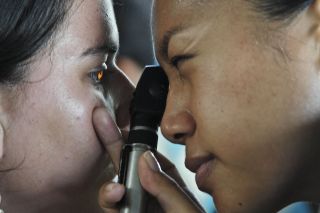
Additionally, it would be ideal to include an ophthalmic examination as part of the AN treatment process. An eye examination could help determine whether patients with AN have any current or potential eye concerns, which might protect them against long-term changes to their eyes.
References
1. Gaudiani JL, Braverman JM, Mascolo M, et al. Lagophthalmos in severe anorexia: A case series. Arch Ophthalmol. 2012;130(7):928–930. doi:10.1001/archophthalmol.2011.2515.
2. Oudman E, Wijnia JW, Oey MJ, et al. Preventing Wernicke's encephalopathy in anorexia nervosa: A systematic review. Psychiat Clin Neuros. 2018;72(10):774–779.
3. Winston AP, Jamieson CP, Madira W, et al. Prevalence of thiamin deficiency in anorexia nervosa. Int J Eat Disorder. 2000;28:451–454.
4. Moschos MM, Gonidakis F, Varsou E, et al. Anatomical and functional impairment of the retina and optic nerve in patients with anorexia nervosa without vision loss. Br J Ophthalmol. 2010.
5. Mroczkowski MM, Redgrave GW, Miller NR, et al. Reversible vision loss secondary to malnutrition in a woman with severe anorexia nervosa, purging type, and alcohol abuse. Int J Eat Disord. 2011;44:281–283.
6. Roda M, di Geronimo N, Pellegrini M, et al. Nutritional optic neuropathies: state of the art and emerging evidences. Nutrients. 2020;12(09):2653. https://doi.org/10.3390/nu12092653.


