


Once the Emperor gave the order, Gulliver tells us, it took 600 Lilliputian beds to make a bed of sufficient “breadth and depth” for him (p. 37) and 300 tailors to make him “a suit of clothing after the fashion of the country. (p. 39, Jonathan Swift, Gulliver's Travels, 1726) Many of the images of Gulliver seem to indicate he was strong and muscular and in good shape.
The careful measurements calculated by the Lilliputians, though, tell us nothing about Gulliver's body composition, namely how much of his body was fat and how much was fat-free.
We have come a long way from the tape measuring days of the Lilliputians. What have we learned since the 18th century?
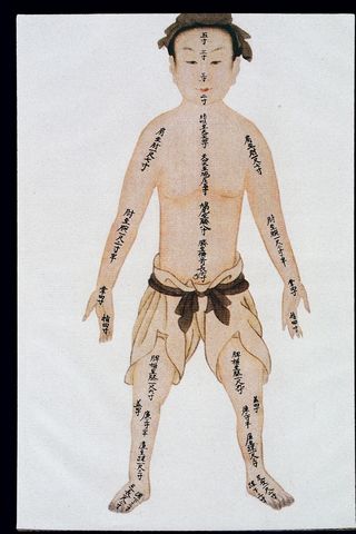
Over the years, researchers have devised many methods to assess body composition, not only to study obesity, but also to study the effects of malnutrition, weight loss following bariatric surgery, altered states of hydration, muscle wasting (sarcopenia) due to old age or to neurological disorders such as Parkinson's or Alzheimer's, cachexia due to chronic diseases such as cancer or HIV, and osteopenia and osteoporosis (Lemos and Gallagher, Current Opinion in Endocrinology, Diabetes and Obesity, 2017; Allison and colleagues in Affuso et al, PLoS One, 2018)
Any method for measuring body composition must consider time, cost, (equipment and personnel), radiation exposure, ease of use, and accuracy. Errors of measurement can be due either to instrument error or to observer error: there is obviously a need for quality control, including training of technicians and calibration of devices. Sometimes, there has to be cross-validation with a subset of a population for reference because body composition can vary by sex, age, and racial or ethnic groups. Even hydration status can affect measurements. (Heymsfield, Allison et al, in Bray and Bouchard, editors, Handbook of Obesity, 2nd edition, 2004, pp. 33-79; Andreoli et al, European Journal of Radiology, 2016)
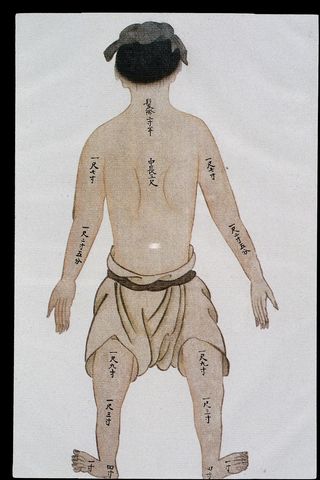
Anthropometry is the least expensive and most widely used method and includes measurement of weight, height, skinfold thickness, and circumferences of body parts such as waist, hips, arms, and thighs. These are considered "proxy" measurements because they don't directly correspond to body composition. (Heymsfield, Allison et al, 2004) Body Mass Index (BMI), the ratio of weight in kilograms divided by height in meters,2 is used conventionally to classify obesity, with a BMI greater or equal to 30 kg/m2 considered obese. But BMI is “not perfectly correlated with body fatness,” and those with the same BMI may have different proportions of fat. (Heymsfield, Allison, et al, 2004) Changes in body weight, for example, can reflect changes in muscle, fat, or water. Further, BMI does not indicate where the fat is located, and it does not differentiate between fat and fat-free mass. (Madden and Smith, Journal of Human Nutrition and Dietetics, 2016)
Obesity is often then “misclassified” when BMI is used to evaluate body fatness (Allison and colleagues, in Affuso et al, 2018) Sometimes, people can be weight-stable while gaining fat and losing muscle. (Prado et al, American Journal of Clinical Nutrition, 2014) For example, as we age, we tend to lose muscle and gain fat, and the mechanisms leading to increased fat deposition as we age are not fully understood. (Gallagher, Heymsfield et al, American Journal of Clinical Nutrition, 2000.) Further, BMI can be inaccurate in muscular athletes, very tall people, or in the elderly with decreased muscle mass (sarcopenia) typical of aging. Gallagher, Heymsfield et al (2000) have collected data to try to establish body fat ranges for optimum health and those ranges directly associated with morbidity and mortality. They found that a "universal set of percentages" is not easily established due to differences in sex, age, ethnicity, and race. BMI cutoffs for risk in Asian populations, for example, are lower than for other races and are even different among different subgroups of Asians. (Nishida et al, European Journal of Clinical Nutrition, 2010)
Assessment of skinfold thickness by calipers measures subcutaneous fat, or "fat patterning," in different areas of the body such as the triceps and subscapular areas, but it is not useful for determining the amount of the dangerous intra-abdominal adipose tissue. Further, equations are "population-specific" and must be cross-validated in sub-populations and often can be very difficult to obtain in very obese patients. (Heymsfield, Allison, et al, 2004)

Circumference measurements (e.g. waist, hips, thighs) are commonly used but they, too, have their own difficulties. For example, a waist has been defined inconsistently by researchers, including using different anatomical landmarks (e.g., the umbilicus or lower margin of the ribs.) Likewise, hip measurements have used different landmarks. (Heymsfield, Allison et al, 2004) A waist circumference, as a surrogate marker of abdominal fat, of greater than 40 inches in men and greater than 35 inches in women, is considered a risk factor for cardiovascular disease. (Klein et al, Diabetes Care, 2007) Many investigators use the waist-to-hip ratio, with a predictor of metabolic disturbances when the ratio is greater than .95 in men and .80 in women, (Heymsfield, Allison et al, 2004), but even this ratio varies among researchers. Karastergiou (Biology of Sex Differences, 2012), for example, used greater than .85 for women in his studies. Further, a waist is often difficult to determine in the very obese, and it may reflect bone and muscle as well as adipose tissue.

These anthropometric measurements continue in use but there are far more sophisticated methods for assessing body composition, including underwater weighing, which is accurate, especially in very obese who cannot be measured easily, but requires subjects to be submerged in water and hold their breath during the process. It is obviously not practical for frail, elderly people, very young children, or those with cardio-pulmonary difficulties. (Heymsfield, Allison, et al, 2004) Another method uses the principle of air displacement inside a sealed chamber, the BOD POD or for infants, the PEA POD. It is reasonably accurate but hydration status may affect results (Lemos and Gallagher, 2017), and the machine is expensive, not readily available throughout the country, and not easily transportable. (Heymsfield, Allison et al, 2004)
Another technique uses the electrical conductive properties of the body--bioimpedance (BIA). It is portable and can be used for field work, but results can vary with posture, hydration status, time of day, and even menstrual cycle, (Heymsfield, Allison et al, 2004) and it is not as accurate as other techniques and suffers from the need to validate in specific populations. (Lemos and Gallagher, 2017; Andreoli et al, 2016)


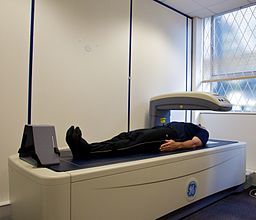
The most commonly used technique for accurate assessment of body composition and the "gold standard" is the Dual Energy Absorptiometry (DXA.) This is the same device that measures bone density to assess osteopenia and osteoporosis. Because it involves radiation, albeit a small dose, it cannot be used in pregnant women, and many parents may not want to expose their children to the radiation. The unit is expensive, must be calibrated regularly, and requires a certified radiology technician.
Over the past 30 years, the scan field and table have been developed to accommodate people of different heights and widths, but if patients are too tall, they may require half-body scans or two scans, and some very obese patients will still not fit on the scanning table. (Silva et al, European Journal of Clinical Nutrition, 2013; Shepherd et al, Bone, 2017)
Other means of assessing body composition include imaging techniques such as MRI or CT scans. An MRI does not require radiation exposure and is considered most valuable for clinical studies as it can assess both regional and depot-specific adipose tissue changes over time. Its disadvantages include its high cost, need for special software that requires considerable training, and difficulties for claustrophobic patients. (Heymsfield, Allison et al, 2004) CT scans are also exceptionally accurate in assessing both subcutaneous and visceral fat, but they require radiation exposure and special software. (Andreoli et al, 2016. See this reference for an excellent chart that compares the pros and cons of each technique.)
Allison and colleagues (Affuso et al, 2018) have developed a new technique to measure the percentage of body fat using two-dimensional digital camera images. Using computer algorithms validated by comparison to DXA, they are in the process of creating norms for subgroups of populations. Their method, of taking three photographs (front, back, and side) is inexpensive, requires minimal training, and no specialized equipment other than a digital camera. It has proven statistically better than assessment by BMI and demographic information and is clearly much more practical for large-scale epidemiological studies than the more sophisticated DXA, MRI, and CT scans.
Bottom Line: Many techniques are now available to measure body composition: anthropometry (e.g., BMI, skinfolds, circumferences of waist, hip, thighs, etc and their ratios.); water displacement; air displacement (BOD POD); BIA: DXA; MRI; CT; and even digital photographs. Each technique has its pros and cons, and none is perfectly "made to measure" everyone, though DXA is today considered the "gold standard" and most commonly used.
Accurate assessment of body composition is useful for evaluation of chronic wasting conditions, such as due to neurological diseases, malnutrition, cancer, or AIDS, or for a work-up for osteopenia or osteoporosis. Importantly, it is an essential part of an evaluation of weight, particularly because where fat accumulates (i.e., whether subcutaneous or visceral) may have serious consequences for health. For a review of these consequences, see my previous blog, Location, Location, Location: The Distribution of Fat.