Self-Control
Neurobiology of Self-Control in Dogs
New brain imaging study shows brain region for canine impulse control.
Posted April 17, 2016
I am a dog-lover, but I will also be the first to admit that my dogs exhibit a sometimes embarrassing lack of impulse control and do things they are not supposed to. Cato, the four year-old plotthound, has not grown out of his habit of chewing the crotches out of underwear. Callie, the seven year-old terrier, still counter-surfs. And Thor, my father-in-law’s big shepherd-mix, who spends time at our house, punched out a pane of glass in the front-door in response to the doorbell (now disconnected). Given that dogs’ natural habitat is with humans, such lapses in behavior can be considered failures of self-control.
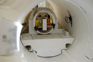
Since 2012, my research group and others have been advancing the field of neuroimaging in awake, unrestrained dogs. The idea is relatively simple: using positive reinforcement, train dogs to go in an MRI and remain still. Once a crazy idea, the technique is increasingly being used around the world to understand how the canine brain functions.
Because the dog is not supposed to move during scanning, researchers have adopted a passive approach. Stimuli are simply presented to the dog, and the resultant brain response is recorded. By measuring which parts of the dog’s brain respond to the stimuli, we can begin to put together a map of how their brains work. Typical stimuli have included hand signals, odors, sounds, and pictures of faces projected on a screen.
Passive responses to stimuli tell us a lot about the organization of perceptual systems, but we would also like to know how these affect behavior. But because of the requirement to remain motionless, we can’t ask a dog to do something in the scanner. The study of behavioral systems in the MRI seemed unattainable. Until now.
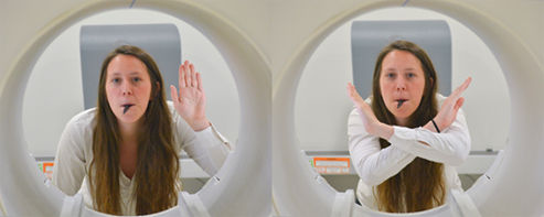
To study self-control, we trained our Atlanta cohort of MRI-dogs on a psychological test commonly used with children. It is called the Go-NoGo test. We taught the dogs to nosepoke a target in response to a whistle.

This was the “Go” condition. This was surprisingly easy to train. Once the dogs had this down, we then introduced the “NoGo” condition. This was a new hand signal in which the arms were raised in an ‘X.’ This meant, “Don’t nosepoke, even when you hear the whistle.” The NoGo condition was harder for the dogs to learn and took 2-4 months of training.

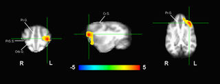
When the dogs were 80% accurate, we had them perform the task in the MRI. Because there was no movement in the NoGo condition, we could capture activity in the brain and see what regions came online to inhibit the tendency to nosepoke. In humans, a region of the prefrontal cortex has been consistently found to be associated with this type of response inhibition. Moreover, the magnitude of this response correlates with other aspects of self-control. A study in 2011 found that the prefrontal response on the Go-NoGo task was correlated with how well people did on the famous “marshmallow test.”

Dogs do not have big frontal lobes, even after accounting for their relatively small brains. Whatever amount of self-control they have must be eked out of a small piece of brain real estate. Until our MRI study, nobody even knew where in the dog’s brain this occurred. When we compared the brain response in the NoGo condition to a neutral condition in which the dog didn’t hear the whistle, we found activation in a small region of the left frontal cortex – very similar to that found in humans. Importantly, the dogs who made fewer errors on the test had more activation in this region, suggesting a direct link between the level of self-control and actual behavior.
We even tested the dogs on a different test of self-control with something called the “A not B” test. This is an old experiment, originally conceived by Jean Piaget as a test of object permanence in human infants. In the A not B test, a dog watches a human place a piece of food in one of three buckets (the A location). She is then released to retrieve the food. This is done three times in the same location, and then, in full view of the dog, the food is moved to a new bucket (the B location). Because the dog has developed a tendency to go one way, they must now override it and go to the new bucket. Sounds simple, but children younger than 9 months can’t do it, and dogs vary widely in their performance.
Interestingly, we found that dogs who did better on the Go-NoGo test did better on the A not B test. This means that there is consistency across different tests of self-control, but dogs vary in their ability to do them. We think this has to do with their underlying brain function.
Why is this important?
My dogs’ lack of impulse control, while sometimes amusing and occasionally damaging, is a nuisance. But the most severe failure of impulse control in dogs is when they bite. The CDC estimates that over 4 million dog bites occur each year, with a disproportionate number occurring in children. Of course there are different reasons why dogs bite, and one can argue whether these represent failures of impulse control, but the end result is a significant morbidity for humans and often the death of the dog.
Our results point to a key location in the dog’s brain. By understanding how this region inhibits responses, we may soon learn why some dogs are better at it than others and potentially ways to improve its function through training.


