Neuroscience
Surfing Brainwaves With EEG
A picture is worth a thousand words. An EEG is worth even more.
Posted May 31, 2017 Reviewed by Matt Huston

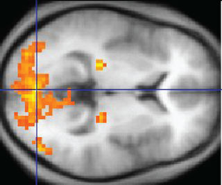
Pictures are powerful tools for illustrating quantitative data and capturing public interest. Each year, NASA releases many beautiful images of Martian dunes and distant nebulae, which help win public funding. Likewise, when it comes to grabbing headlines and commanding public attention, studies of functional brain activity often do best when they beautifully illustrate said activity as colorful pixels dancing on the convoluted surface of the cerebral cortex.

You've probably heard of functional magnetic resonance imaging, or fMRI, the technology that produces stunning brain scans seen on the evening news. However, these images are missing a critical dimension: time. Brain activity takes place on a millisecond timescale, yet fMRI captures this activity at a rate of about one full brain scan per second. This is rather akin to watching a movie filmed at a rate of one frame per second.
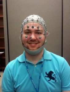
EEG, short for electroencephalogram, is a comparatively old technology, first introduced by Hans Berger in 1929, in which electrodes placed on the scalp record the brain’s electrical activity (“brainwaves”) with millisecond resolution. By taking thousands of samples per second, EEG captures the time course of subsecond neural responses to stimuli. Furthermore, EEG is a direct measure of brain activity taking place at parts of neurons called synapses and dendrites, whereas fMRI instead measures metabolic activity as a proxy for synaptic activity and neuronal firing.

While one might naively assume EEG to be a primitive technology given its relative antiquity and inability to produce sexy pictures, modern computers yield tremendous amounts of information from EEG recordings. Though originally recorded with a moving pen in seismograph-like fashion, digitized EEG data in the ’80s allowed for mathematical transformations of EEG recordings to show a spectrum of brain oscillations. Different brain oscillations or "brainwaves" are associated with different cognitive tasks and brain processes. The alpha rhythm, for instance, is associated with neural “idling” in the resting brain; the gamma rhythm is associated with cognition and coordination of brain regions processing different aspects of the same information.
With the advent of faster computers, mathematics from the fields of chaos theory and information theory has recently been used to quantify the complexity of EEG recordings. These metrics offer promise for identifying biomarkers (objective, quantifiable signatures) of brain disorders such as schizophrenia, autism, and Alzheimer’s. In normal clinical practice, EEG has been used for decades to diagnose epilepsy and sleep disorders; it is also an invaluable tool for observing comas and monitoring depth of anesthesia. Being cheap and highly portable, EEG tests are easier to administer than MRI brain scans and more practical for many purposes.
EEG and fMRI are both useful tools for measuring functional brain activity, each with its own strengths and weaknesses. Being recorded from the scalp, EEG has poor spatial localization but addresses what questions with its high temporal (i.e., time) resolution. By contrast, fMRI has excellent spatial localization for addressing where questions, but often lacks the temporal resolution to tell us what is going on in the brain. For this reason, fMRI is most appropriate when a researcher wishes to address an anatomical hypothesis (e.g., what brain region does this take place in?). The brain treads a delicate balance between functional segregation and integration: cognitive tasks are partially localized to specific brain regions and partially distributed across the whole brain. EEG and fMRI are both appropriate for testing different hypotheses in different contexts.
This post was adapted from a previous post by Joel on Knowing Neurons.
References
Mulert, Christoph, and Louis Lemieux, eds. EEG-fMRI: physiological basis, technique, and applications. Springer Science & Business Media, 2009.


