Pregnancy
When a Fetus Turns to Stone
Rare “stone babies” reveal how a woman’s body can cope with a dead fetus.
Posted March 5, 2020 Reviewed by Kaja Perina
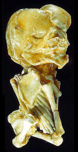
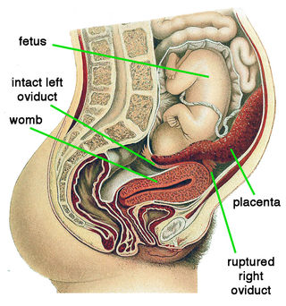
My blog post "Ectopic Embryos: Pregnancy in the Wrong Place" discussed unusual cases in which human pregnancies begin outside the womb. Occurring in just over 1% of pregnancies, this is a significant cause of fetal and maternal death even with good health care. An ectopic pregnancy is almost always due to a fertilized egg attaching inside an oviduct on its way to the womb (tubal pregnancy). Exceptionally — once in about 10,000 pregnancies — the fertilized egg does not even arrive in the oviduct and attaches to the outside of the womb, an ovary or the intestines (abdominal pregnancy). Surgery might seem to be the only option to save a woman with a fetus developing in the wrong place. Remarkably, however, women occasionally survive for decades with a dead fetus in the abdomen, literally petrified as a “stone baby” (lithopaedion).
How a stone baby forms
A stone baby can develop only after three months of pregnancy. Before that, an embryo or early fetus is small enough for the woman’s body to resorb. With a bigger fetus, however, the mother can instead mobilize a natural response to foreign bodies: depositing calcium salts to isolate the decaying tissue and forestall infection. This occurs mainly with ectopic pregnancies. In a landmark 1881 review — followed by physicians ever since — Friedrich Küchenmeister distinguished three types of calcification: a stone sheath when the membranes surrounding the fetus remain intact and become petrified, a stone baby when expulsion into the abdominal cavity ruptures the membranes and the fetus itself is petrified, and a stone sheath baby when both membranes and fetus are calcified.
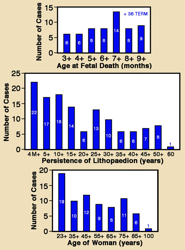
Another seminal paper by Daniel Tien (1949) thoroughly surveyed stone babies, reviewing 247 previous cases. Although Küchenmeister’s three types of lithopaedion were almost equally common, true stone babies predominated. Two-thirds of adequately documented cases originated with tubal pregnancies, while only a tenth were attributed to pregnancy in the womb. Tien confirmed that stone babies develop only between the fourth month of pregnancy and term, on average during month 7. Stone babies were discovered with women aged between 23 and 100 years (average 55), over half after menopause. Before discovery, stone babies had been present for between 4 months and 60 years (average 22 years). Nine women unwittingly carried stone babies for over 50 years prior to detection! In several cases, women uneventfully conceived and gave birth while carrying a lithopaedion.
Recent case reports
Following Tien’s review, medical journals have published many case reports of stone babies, almost doubling their number. One well-documented, typical report by Chia-Ming Chang and colleagues (2001) described the case of a 76-year-old woman hospitalized for surgery because of a tumour on the neck of her womb. While totally removing her womb, the surgeon incidentally discovered a stone baby in her abdomen. Her medical history was unexceptional and laboratory test results were normal. The patient remembered suffering severe abdominal pain some 50 years previously. At the time, after palpating her pelvic region, her physician had diagnosed a benign tumour.
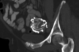
A widely-cited 1999 report by Craig Frayer and Milo Hibbert described finding a lithopaedion in a 67-year-old woman, following an abdominal pregnancy, that remained undetected for 37 years. Acute abdominal pain drove the woman to seek help at a hospital emergency department, where X-rays revealed a fetal skeleton between her pelvis and ribcage. A pregnancy test was negative and pelvic examination indicated a normal post-menopausal womb. The patient had been diagnosed as having a "missed pregnancy” 37 years ago, but refused intervention then and had no adverse effects thereafter. In the interim, she experienced a healthy, uneventful pregnancy ending with a normal vaginal birth.
Early history of stone babies
A 1996 paper by medical historian Jan Bondeson reported on the earliest adequately documented case of a stone baby, discovered in 1582 during an autopsy conducted on a 68-year-old woman in Sens, France. She had carried the lithopaedion in her abdomen for 28 years. Propelled by morbid curiosity, the Sens stone baby became a much-coveted relic, changing owners across Europe repeatedly in the 1600s. Damaged by careless handling, it ended its travels in Copenhagen but subsequently disappeared without trace.
The first written record of an abdominal pregnancy dates back over 1000 years to the renowned 10th Century surgeon Abu Qasim Al-Zahrawi (widely known as Abulcasis), who observed parts of a fetus being expelled from a woman’s belly near the navel.
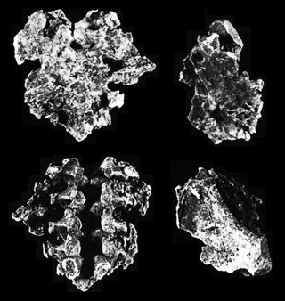
In 1993, Bruce Rothschild and colleagues reported far earlier archaeological evidence of a stone sheath lithopedion from a 3100-year-old hunter-gatherer sinkhole burial site on the Edwards Plateau in Texas, USA. Four fragments were found with isolated skeletal elements — notably vertebrae from the backbone — fused to a shield-like portion of structureless calcified membrane. Size of the vertebrae indicated that the fetus died around the 8th month of pregnancy.
Special cases
The literature also includes extremely rare accounts involving twins, starting with a 1952 paper by Keeling Roberts. Some 18 months after giving birth to a child, a 29-year-old woman sought medical attention because of menstrual irregularity. Examination indicated that she was 5-6 weeks pregnant. After another two months, the woman experienced pain low down on her right side and examination revealed that the womb extended almost up to her navel. At 30 weeks of pregnancy, a single fetal heartbeat was detected and an X-ray confirmed presence of a single normal fetus. The patient herself was seemingly healthy. Examination a month later revealed that the fetus was in breech presentation, which was easily corrected. When the patient was two weeks overdue, birth was induced, and labour began immediately. Once the patient was fully dilated, an almost completely calcified fetus was found wrapped around the back of the emerging baby’s head. An area of dead tissue on the placenta indicated the location of the umbilical cord leading to the lithopedion, which had died about halfway through pregnancy.
In two recently reported cases, calcification affected both fetuses in twin pregnancies. A 2006 paper by Puneet Misra and colleagues reported findings for a 45-year-old woman who attended their hospital in Ganjam District, Orissa, India, complaining of lower abdominal pain. X-rays revealed the presence of calcified Siamese twins (two heads on a single body) that had escaped diagnosis for 18 years. Thigh bone length measured with ultrasound indicated that the two-headed fetus had died after seven months of pregnancy.
A year later, Jagmohan Mishra and colleagues reported a second case of calcified twins with a 40-year-old patient at a hospital in Burla, Orissa, India. Eight years previously she had a missed abortion in the fifth month of pregnancy. After being admitted to hospital in 2005 because of a week-long acute intestinal obstruction, X-rays showed two rounded dense masses on left and right sides of the lower abdomen. During emergency laparotomy, a section of strangulated intestine attached to a solid mass behind the left ovary and a similar solid mass from the right side of the pelvic cavity were removed. Each mass contained a 5-month-old calcified fetus surrounded by a tough envelope.
Surviving abdominal pregnancies
Most abdominal pregnancies originate when an embryo begins to develop in an oviduct. When the fetus reaches a critical size, the oviduct bursts and spills it into the abdomen (secondary abdominal pregnancy). Primary abdominal pregnancies with the embryo developing from the outset in the abdominal cavity are very rare. Amazingly, women occasionally survive abdominal pregnancy without surgery when calcification converts the dead fetus into a stone baby. It can then remain undetected for decades until discovered incidentally during a medical examination/operation for other reasons or at autopsy. Palpation in the pelvic region, X-rays, CT-scans and MRI images can all reveal the presence of an abdominal mass. So one really has to ask why unsuspected cases are still being discovered even in countries with state-of-the-art health facilities!
Fortunately, women carrying a lithopaedion commonly show no distinctive side-effects. They may experience a sensation of weight in the abdomen, pelvic pain or compression of organs (especially the urinary bladder or rectum). Stone babies will hopefully become even rarer in the future with advances in diagnosis and timely surgical intervention. But increasingly frequent inflammatory pelvic disease and oviduct surgery are reportedly increasing the occurrence of ectopic pregnancies.
Acknowledgment: I am very grateful to Bruce Rothschild for kindly allowing me to use a modified version of the figure from his co-authored 1993 paper.
References
Bondeson, J. (1996) The earliest known case of a lithopaedion. Journal of the Royal Society of Medicine 89:13-18.
Chang, C.-M., Yu, K.-J., Lin, J.-J., Sheu, M.-H. & Chang, C-Y. (2001) Lithopedion case report. Chinese Medical Journal (Taipei) 64:369-372.
Frayer, C.A. & Hibbert, M.L. (1999) Abdominal pregnancy in a 67-year-old woman undetected for 37 years. A case report. Journal of Reproductive Medicine 44:633-635.
Küchenmeister, F. (1881) Ueber Lithopädien. Archiv für Gynäkologie 17:153-159.
Mishra, J.M., Behera, T.K., Panda, B.K. & Sarangi, K. (2007) Twin lithopaedions: a rare entity. Singapore Medical Journal 8:866-868.
Misra, P., Bastia, B.K., Deep, N., Rao, M. & Jena, S.K. (2006) Lithopaedion evolution of an abdominal siamese twin pregnancy — a rare case report. Indian Journal of Radiology & Imaging 16:813-814.
Roberts, J.K. (1952) A case of lithopedion in twins. British Medical Journal 2:130.
Rothschild, B.M., Rothschild, C. & Bement, L.C. (1993) Three-millennium antiquity of the lithokelyphos variety of lithopedion. American Journal of Obstetrics and Gynecology 169:140-141.
Santoro, G., Laganà, A.S., Sturlese, E., Giacobbe, V., Retto, A. & Palmara, V. (2014) Developmental and clinical overview of lithopaidion. Gynecologic & Obstetric Investigation 78:213-223.
Tien, D.S.P. (1949) Lithopedion: general discussion and case report. Chinese Medical Journal 67:451-460.


