Neuroscience
The Eyes Have It
Unraveling the brain by tugging on a retinal thread
Posted October 8, 2012
I recently attended a fantastic lecture by physicist-turned-neuroscientist Sebastian Seung of MIT, held as part of the Bio-X seminar series here at Stanford. The lecture showcased some initial results concerning the structure and function of the retina, and in particular on the mechanisms behind the cells that allow us to perceive motion, which, I think you’ll agree, is a pretty important ability to have. Now you might wonder why I’m writing about eyes in a blog about the brain, but as my ophthalmologist wife likes to remind me, the retina is part of the brain—the part of the body where the brain has pushed itself outside of the skull to get a look at the world—and so she actually examines far more brains than I do! The work demonstrates the potential of the new science of connectomics to move neuroscience forward—and as an added bonus, Seung’s research platform offers the opportunity for you to actually do some science (more on this below), and help solidify the preliminary models I’ll describe. Plus, the pictures are beautiful (you can see more at the eyewire.org wiki, which is where I got most of these here).
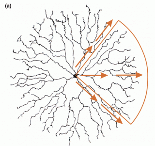
The story starts with a retinal cell called the starburst amacrine cell (SAC). This is a pretty interesting neuron in its own right. First, it has no axon, just the starburst of dendrites—those splayed threads you see here in figure 1. SACs secrete two different kinds of neurotransmitter molecules—acetylcholine (ACh) and GABA—that is, both a primarily exitatory and a primarily inhibitory neurotransmitter, which is pretty unusual. Moreover, each dendrite is relatively independent of the others, such that each sends a signal when it detects motion that matches the direction of the dendrite itself (figure 2; Masland 2005).

Figure 1: Starburst Amacrine Cell

Figure 2: SAC directional selectivity
So this one cell is able to detect motion in any direction, and sends a signal from just the dendrite that indicates that direction. Interestingly, one of the main mechanisms that permits this sort of single-cell multifunctionality is the pattern of connections to and between these SACs, which interacts with cell morphology in just the right way. Loosely speaking, the inputs to each individual dendrite are arranged so that light-detecting bipolar cells connect serially along the dendrites, in an arrangement in which a neighboring bipolar cell makes a neighboring synapse on a dendrite, making it more likely that motion in that direction will cause signals that reinforce along that dendrite. In addition, neighboring SACs selectively reinforce and inhibit one another, so that dendrites going in the same direction reinforce each other, while dendrites going in different directions inhibit one another. The resulting directional selectivity of the dendritic signal is so useful that these cells are used in multiple higher-level functions, including motion perception and also the control of eye movements. These details are interesting in their own right, I think, but also underline some important principles of structure-function relationships in the brain: 1) even individual cells can be multi-functional in various ways, doing different things at different times, and having multiple higher-level uses; 2) the source of neural function isn’t just a matter of the intrinsic, local properties of the cell, but of global properties of the network; and 3) physical structure matters. If these dendrites weren’t arrayed spatially in this way, the cell couldn’t have the functional properties it does.

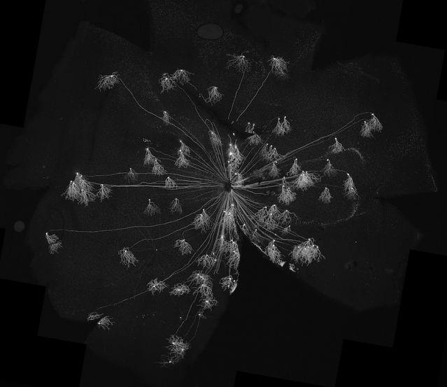
Figure 3: JAM-B cells in the mouse retina
This last point is further underlined by an intriguing hypothesis for the mechanism of directional selectivity of the so called JAM-B cells. The gorgeous figure 3 shows a number of these cells in mouse retina, with the long axons traveling along the back of the eye to the optic nerve in the center, and the dendritic arbors all pointing in the same direction.
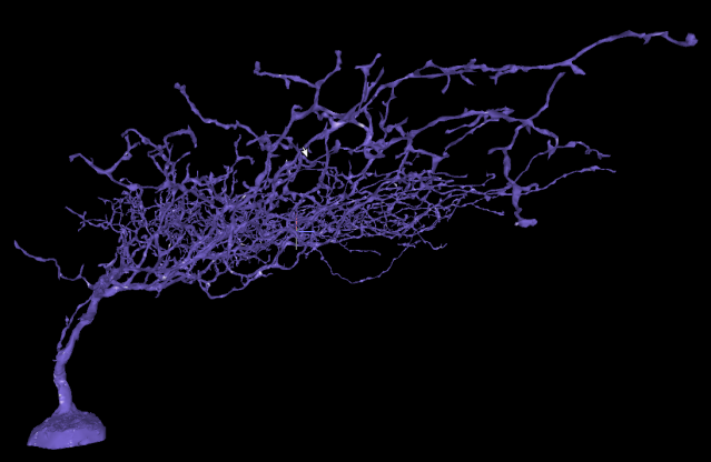
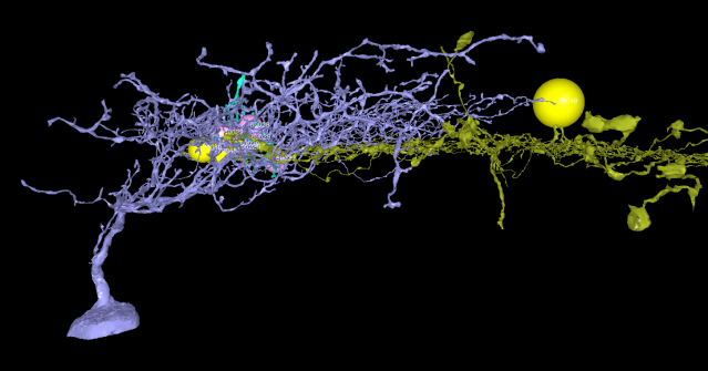
JAM-B cells respond only to upward motion (remember the image on the retina is upside-down), and the question is, how? Well, the asymmetry of the dendritic arbors gives these cells a distinctive depth profile, as shown in figure 4. Seung’s hypothesis is that an important part of the mechanism for directional selectivity in these cells involves inhibitory synapses from SACs—they will prevent the cell from responding when the direction of motion isn’t up. Now, in fact, SACs make very few connections to JAM-B cells, and so one would think that their influence would be minimal. But here’s the crucial part of the hypothesis: SACs exist in a lower layer of the retina, and so the connections they do make to JAM-B cells will tend to be lower in the dendritic branches, closer to the cell body (yellow cell, figure 5).

Figure 4: JAM-B side view
Thus, the few connections SACs do make to JAM-B cells may be perfectly positioned to inhibit signals traveling to the cell body. Put differently, an important determinant of function in this case may be the relative spatial position of the neurons in the circuit. As important as it is to know that cells are connected, it can also be crucial to know where they are connected. The details of our embodiment matter in sometimes surprising ways.

Figure 5: Spatial relationship between JAM-B cell (purple) and SAC (yellow)
And the work is only just beginning—imagine what other secrets the brain has to whisper in your ear! The good news is you don’t have to just imagine it, you can actually listen for yourself. For although it may seem as if a recurring thesis of this blog is something like: neuroscience is hard! (and the work outlined above certainly cements this basic fact), in this case there is the opportunity for some fun. Because tracing individual neurons in the retina is incredibly time-consuming and expensive, Seung has decided to try and crowdsource part of the work. Hypotheses like those described above are only possible in light of the structural results that connectomics like this can provide. So go to eyewire.org and help unravel the mysteries of the brain by playing the eyewire game. It’s probably less fun than Halo 4, but you’ll need something to do before that’s released—and this way you can say you’re helping to build a brain. By playing Xbox, maybe not so much . . .
Masland, R. H. (2005). The many roles of starburst amacrine cells. TRENDS in Neurosciences, 28(8): 395-6.


